Brain tumors vary in type.
Secondary brain tumors: These are tumors that spread from another part of the body to the brain and can be cancerous, such as tumors of the cells lining the pharynx.
- Primary brain tumors: These are tumors that originate inside the brain and vary in type, including:
Benign tumors, such as hemangiomas, choroid plexus tumors, schwannomas, chondromas, and pituitary adenomas.
Malignant tumors: Squamous cell carcinoma, acoustic neuroma, and pituitary gland tumors.
There are, of course, stages of tumors, but cancerous tumors are more dangerous to humans because they spread more quickly and work to spread cancer to healthy cells. Symptoms usually arise when the tumor is large and can cause headaches, visual and auditory disturbances, loss of balance, and significant problems for the body.
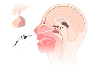
Skull base tumor surgery is one of the most delicate surgical procedures and requires high skill and precision in surgery. Dr. Islam Alajhouri, an assistant professor of neurosurgery and spinal surgery, and holder of a doctorate in skull base tumors and a fellowship in brain endoscopy from the University of Greifswald, diagnoses the tumor by reviewing the patient’s medical history, conducting hearing and visual tests, balance tests, and performing magnetic resonance imaging, computed tomography scans, and laboratory tests.
The procedure is performed through the nose or mouth using surgical endoscopes through the nostril openings. The tumor is removed, and a sample is taken to confirm the diagnosis.
Endoscopic brain ventricle surgeries
The ventricles of the brain are cavities connected to each other inside the brain. They produce, transport and displace cerebrospinal fluid that floods the central nervous system. This helps keep the brain active. The brain ventricle consists of 4 ventricles that form a connected network of cavities.
The lateral ventricles are located on the left and right sides of the brain, and their size increases with increasing age, and may be asymmetrical, and cerebrospinal fluid is produced
The third ventricle is a slit located in the midline region of the brain and is connected to both the lateral ventricles and the fourth ventricle and contains cerebrospinal fluid
The fourth ventricle is located at the top of the medulla and serves to protect the brain from trauma, helping to form the central canal that runs along the spinal cord.
It is a disease of the ventricles of the brain
Enlargement of the brain ventricles or obstruction of the brain ventricles, which is a significant increase in the size of the ventricle as a result of inflation of the cerebrospinal fluid in certain areas, which pushes the ventricles of the brain to grow larger due to hydrocephalus caused by blockage of the passage between the ventricles and obstructing the flow of cerebrospinal fluid through the ventricles of the brain. This operation is considered an alternative option to the shunt operation for the treatment of hydrocephalus
Meningitis is caused by a bacterial or viral infection and is a swelling of the tissues surrounding the brain and spinal cord
Brain hemorrhage is a stroke in the form of bleeding both inside the brain due to high blood pressure and abnormally weak and dilated blood vessels.
This type of surgery requires precision and special skills from an experienced surgeon, and he has successful experiences in those operations that are available in Dr.
Endoscopic brain surgeries
It is a surgical operation that does not require a hole in the skull, but small holes are made in the skull or through the nose, and these holes are small, ranging between 5 mm and 15 mm, through which a telescope and surgical tools are inserted, and it includes the presence of a camera. Endoscopic brain surgeries are very accurate and careful, with the ability to magnify up to 9 times. Endoscopic brain surgeries are done to remove tumors, vascular deformities, aneurysms in the brain, and cases of hydrocephalus.
Advantages of endoscopic brain surgeries
It is characterized by high accuracy during the procedure.
Less painful than traditional surgeries.
The patient's return to daily life faster due to the smaller size of the wound.
The endoscope enlarges the image, which facilitates access to the surgical site accurately and increases the chances of success of the operation and recovery.
The risk of injury to healthy tissues is minimal, which prevents complications and reduces wound inflammation and adhesions that may occur.
Not losing a lot of blood during the operation.
Preserves brain tissue and protects it from laceration by making small openings.