
Microscopic surgery in the brain
It is a minimally invasive surgical procedure that involves using specialized surgical micro-tools and precision instruments to repair complex structures such as blood vessels and nerves, with a limited and precise penetration. It is performed through small incisions based on the location of the problem in the brain.
Surgical microscope is considered one of the best and safest techniques for removing brain tumors, as Dr. Islam Al-Ajhuri can safely remove as much of the tumor as possible without damaging the surrounding brain tissue.
Neuronavigation systems also assist in brain and nerve surgeries by providing real-time guidance during the procedure. This ability increases the accuracy and safety of brain and spinal cord surgeries.
Brain tumor removal and vascular surgery.
Brain tumors.
It is an irregular growth in brain cells ranging in size from small tumors to very large tumors. The tumor may be small, but it has a significant impact and symptoms. While other types of brain tumors grow to a large size before they are discovered because of its presence in a less active form than others, so it must be diagnosed
Excision of brain tumors and vascular surgery
Rapidly.
Types of brain tumors
Benign tumors:
It is the appearance of a mass of cells growing abnormally in this area, and this growth is slow and the benign tumor cannot invade other organs of the body, but the problem with the benign tumor is that it may press on vital blood vessels or nerves inside the brain.
Malignant tumors:
Abnormal cells grow and spread at a high speed and move to the surrounding tissues, and they do not die unlike normal cells.
It varies according to its influence
Primary tumors: They appear as a result of a defect in the brain cells, so they begin to divide unaccounted for, and a tumor appears.
Secondary tumors: It is the appearance of a tumor in the brain as a result of the spread of cancer cells that initially affected another organ, such as the lung or kidney.

Brain tumor removal
It is a surgery in which surgery is performed to remove the tumor located in any part of the brain and is considered the best treatment method for benign tumors without causing any neurological impairment.
Brain cancer is one of the most common types and is characterized by its rapid growth, causing pressure on the brain tissue, which in turn leads to its damage.
Aneurysm
It occurs due to a weakness in the wall of the blood vessel located inside the brain, as that area of the blood vessels is eroded due to the continuous flow of blood, and the symptoms of aneurysm are characterized by sudden headache, nausea, sensitivity to light, blurred vision, and stiffness in the neck.
Types of aneurysms
Cystic aneurysms: They are more common, as the vessels bulge and look like a dome.
Fusiform aneurysm: This type of aneurysm is rare in the brain, causing a dilated spot within a blood vessel.
Excision of tumors of the base of the skull
The base of the skull is a complex and irregular bony surface on which the brain rests. It contains major and large blood vessels that supply the brain with the basic nutrition needed for it, and the important nerves with their exit passages from the brain.
varied
Tumors of the base of the skull
Depending on the type of tumor and its location inside the base of the skull, whether it is in the front, middle or back.
The patient suffers from headache, facial numbness, difficulty breathing, and facial pain
Diagnostics of skull base tumors
Through brain imaging, a tumor at the base of the skull by CT scan, magnetic resonance, positron emission tomography, and examining the skull bones can be diagnosed by taking a small sample of the tumor at the base of the skull and examining it under a microscope. The sample can be taken using an endoscope that is placed through the nose and paranasal sinuses.
Tumors are removed through the wedge-shaped operation, which is done through the nostrils or through the mouth. The process begins by inserting the end of the endoscope through the nostril and using a microscope. Lighting and magnification are available, so the neurosurgeon, Dr. Surgery and laparoscopy followed, as he dealt with the most sensitive organs of the body, which is the brain, and achieved the highest success rate in these operations.
Acoustic neuroma surgeries
An acoustic neuroma is a noncancerous tumor that usually grows slowly on the main nerve that leads from the inner ear to the brain. The pressure resulting from an acoustic neuroma can cause progressive hearing loss, ringing in the affected ear, unsteadiness or loss of balance, dizziness, numbness of the face, and weakness or loss of muscle movement. The diagnosis is made by means of medical imaging. Magnetic resonance imaging and acoustic neuroma surgery is performed under general anesthesia and includes removal of the tumor through the inner ear.
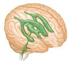
Microscopic surgery of the ventricles of the brain
ventricles of the brain
They are hollow areas within the brain that are filled with cerebrospinal fluid, a clear fluid that surrounds and lines the brain and spinal cord.
Ventricular tumors are a common type of tumors that grow in the lining of the brain, and ependymal tumors lead to tumors that are more invasive and difficult to remove. Therefore, it is necessary to choose the appropriate doctor accurately, depending on his long experience and the success rate of the operations he performed. Among those symptoms are headaches or a change in their pattern that becomes more difficult and severe, and unjustified nausea or vomiting, and they are diagnosed through a clinical examination or magnetic resonance imaging to make a decision on the treatment of an ependymoma patient, taking some considerations, such as The age of the patient, the location of the tumor, its degree, and possible side effects, then a microscope is used to remove the tumor without a major surgical intervention.He is an assistant professor of neurosurgery and spine surgery at Al-Azhar University. He is also a consultant neurosurgeon and spine surgeon at the Police Hospital and a member of the Royal College of Surgeons of England.
Therefore, he is considered one of the best and best surgeons, because he was trained in the best techniques of microscopic surgery, surgical navigator, and endoscope. He also dealt with the most sensitive organs of the body, which is the brain, and achieved the highest success rate in these operations.